 |
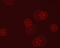
Canberra, Australia (SPX) Feb 10, 2009 CSIRO has patented an improved microscopy method for measuring the shapes and sizes of proteins which could help scientists create new pharmaceuticals that are a better match for the proteins they target. The method, called Differential Aberration Correction (DAC) microscopy, measures distances at the molecular level in two and three dimensions using conventional fluorescence microscopy. A special feature of the new method, written up recently in the Journal of Microscopy, is that it allows scientists to measure proteins in solution, which is how they exist in nature, instead of using coated or crystallised proteins as other techniques do. This is particularly important for rational drug design but has broader life sciences applications. The leader of CSIRO's Biotech Imaging team, Dr Pascal Vallotton, says DAC microscopy measures distances a million times smaller than a tape measure can - in nanometres rather than millimetres. One nanometre is one billionth of a metre. "Just as a tailor measures up a person for a suit, we want to use our technique to measure accurate dimensions of proteins called membrane receptors," Dr Vallotton says. "These proteins sit on cell boundaries, acting as gate-keepers, and they represent a class of biomolecules targetted by over 50 per cent of pharmaceuticals. "Understanding the complex structures of these molecules and how new drugs affect their structure will help drug companies design more effective pharmaceuticals." Cells are often viewed using conventional fluorescence microscopy. However, images obtained this way are inaccurate because light is bent differently for different wavelengths through the microscope. Several universities have attempted complex and onerous hardware-based solutions to try to fix this problem. CSIRO researchers instead used a software-based approach that precisely corrects the distortion at every point in the image. DAC microscopy is an improvement on an older technology, called FRET, which can measure distances from 1-10 nanometres. DAC can measure 1-250 nanometres, giving a more complete picture of drug-membrane receptor interactions. It will complement other techniques like X-ray crystallography. The DAC software was tested using fluorescent polystyrene microspheres only 100 nm across - about one thousandth the width of a hair. "We are currently looking at more samples that require the ability to see single molecules with very high contrast. Results are extremely encouraging and we are starting to explore commercial possibilities," Dr Vallotton says. The DAC software was recently demonstrated in the US and will be presented at the Society for Biomolecular Screening conference in Lilles, France in April. Related Links CSIRO Mathematical and Information Sciences Hospital and Medical News at InternDaily.com
 West Lafayette IN (SPX) Feb 10, 2009
A research center in Purdue's Discovery Park, entering its fifth year of operation, is building on several new research projects for expanding efforts in applying systems-engineering efforts to improve the nation's health-care system. |
|
| The content herein, unless otherwise known to be public domain, are Copyright Space.TV Corporation. AFP and UPI Wire Stories are copyright Agence France-Presse and United Press International. ESA Portal Reports are copyright European Space Agency. All NASA sourced material is public domain. Additional copyrights may apply in whole or part to other bona fide parties. Advertising does not imply endorsement, agreement or approval of any opinions, statements or information provided by Space.TV Corp on any Web page published or hosted by Space.TV Corp. Privacy Statement |